

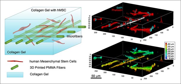
Schematic illustrations of 3D printed fiber: cell-laden collagen gel composites. Right image displays 3D fluorescence and depth-coded of constructed image of fiber: cell-laden collagen gel displaying the location of cell and fibers in different depth of the 3D gel.
Students develop efficient 3D near-field electrospinning for potential tissue engineering
7/3/2017
UNIVERSITY PARK, Pa. — Biomedical engineers at Penn State have demonstrated a simple and innovative method for fabrication of highly organized polymeric fiber structures blended with stem cell loaded collagen gels.
The new technology, 3D near field electrospinning (3DNFES), combines 3D printing and near field electrospinning techniques. 3DNFES is capable of placing single micrometer scale fibers in a predefined spatial organization on multiple substrates. This new method could improve the way nano- and micro fibers have been used in regenerative medicine, such as neural regeneration research, or other applications, such as drug delivery.
Creating fine and porous scaffolds capable of mimicking the natural extracellular matrix (ECM) using polymeric fibers (nano and micron size) is essential for tissue engineering and regenerative medicine. It is fairly difficult to generate organized networks using conventional fiber fabrication methods such as electrospinning.
In a recent paper published in Advanced Healthcare Materials, Justin Brown, associate professor working with Pouria Fattahi, a doctoral student in biomedical engineering, and Jordan T. Dover, an undergraduate in biomedical engineering, describes the novel 3DNFES printer technique for fabrication of pre-patterned polymer fibers.
The development of this new breakthrough, 3DNFES, can fill the gap for fabrication of precise patterned fibers with enormous practical application not only in biomedical areas but also in micro- and nanodevices and electrical and optical applications.
Share this story:
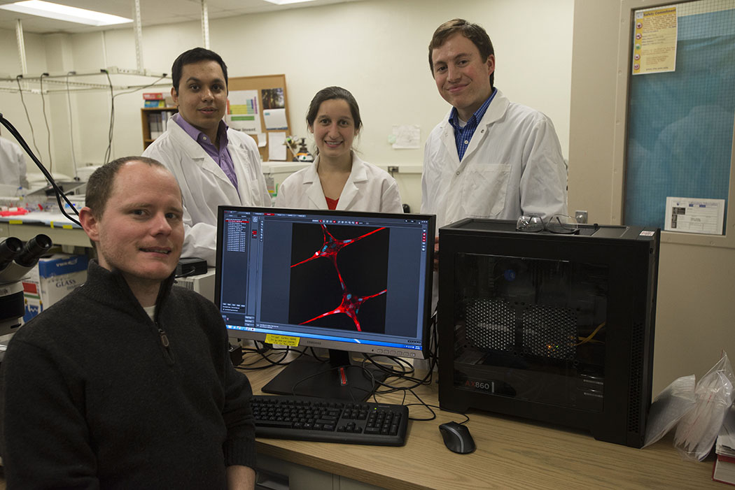
Associate Professor Justin Brown works with students on regenerative medicine and tissue engineering research.